Salter–Harris fracture
| Salter–Harris fractures | |
|---|---|
| Other names: Growth plate fracture[1] | |
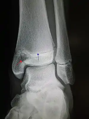 | |
| An X-ray of the left ankle showing a Salter–Harris type III fracture of medial malleolus. Red arrow demonstrates fracture line while the blue arrow marks the growth plate. | |
 | |
| Specialty | Orthopedics |
Salter–Harris fracture is a bone fracture involving the growth plate.[2] It is thus a form of child bone fracture.
It is a common injury found in children, occurring in 15% of childhood long bone fractures.[3] This type of fracture and its classification system is named for Robert B. Salter and William H. Harris who created and published this classification system in 1963.[4]
Types
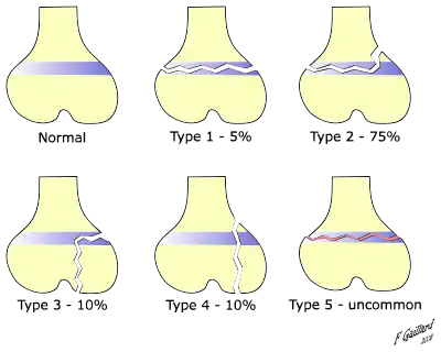
There are nine types of Salter–Harris fractures; types I to V were originally described,[3] and types VI to IX were added subsequently:[5]
- Type I – transverse fracture through the growth plate (also referred to as the "physis"):[6] 6% incidence
- Type II – A fracture through the growth plate and the metaphysis, sparing the epiphysis:[7] 75% incidence, takes approximately 12-90 weeks or more in the spine to heal.[8]
- Type III – A fracture through growth plate and epiphysis, sparing the metaphysis:[9] 8% incidence
- Type IV – A fracture through all three elements of the bone, the growth plate, metaphysis, and epiphysis:[10] 10% incidence
- Type V – A compression fracture of the growth plate (resulting in a decrease in the perceived space between the epiphysis and metaphysis on x-ray):[11] 1% incidence
- Type VI – Injury to the peripheral portion of the physis and a resultant bony bridge formation which may produce an angular deformity (added in 1969 by Mercer Rang)[12]
- Type VII – Isolated injury of the epiphyseal plate (VII–IX added in 1982 by JA Ogden)[13]
- Type VIII – Isolated injury of the metaphysis with possible impairment of endochondral ossification
- Type IX – Injury of the periosteum which may impair intramembranous ossification
.jpg.webp) Salter–Harris I fracture of distal radius.
Salter–Harris I fracture of distal radius. Salter–Harris II fracture of ring finger proximal phalanx.
 Salter–Harris III fracture of big toe proximal phalanx.
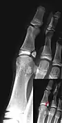 Salter–Harris IV fracture of big toe proximal phalanx.
Mnemonic
The mnemonic "SALTER" can be used to help remember the first five types.[14][15][16]
N.B.: This mnemonic requires the reader to imagine the bones as long bones, with the epiphyses at the base.
- I – S = "Slip (separated or straight across)". Fracture of the cartilage of the physis (growth plate)
- II – A = "Above". The fracture lies above the physis, or "away" from the joint.
- III – L = "Lower". The fracture is below the physis in the epiphysis.
- IV – TE = "Through everything". The fracture is through the metaphysis, physis, and epiphysis.
- V – R = "Rammed (crushed)". The physis has been crushed.
Alternatively, SALTER can be used for the first 6 types, as above but adding Type V — 'E' for 'Everything' or 'Epiphysis' and Type VI — 'R' for 'Ring'.
Prognosis
Fractures in children generally heal relatively fast but may take several weeks to heal.[1] Most growth plate fractures heal without any lasting effects.[1] Rarely, bridging bone may form across the fracture, causing stunted growth and/or curving.[1] In such cases, the bridging bone may need to be surgically removed.[1] A growth plate fracture may also stimulate growth, causing a longer bone than the corresponding bone on the other side.[1] Therefore, the American Academy of Orthopaedic Surgeons recommends regular follow-up for at least a year after a growth plate fracture.[1]
History
Types I to V were described by Robert B Salter and W Robert Harris in 1963,[3] and the rarer types VI to IX were added subsequently.[17]
See also
- Paul Jules Tillaux
- Thurstan Holland sign
References
- 1 2 3 4 5 6 7 "Growth Plate Fractures". orthoinfo.aaos.org, by the American Academy of Orthopaedic Surgeons. Archived from the original on 2018-02-05. Retrieved 2018-02-05. Last Reviewed: October 2014
- ↑ Cepela, Daniel J.; Tartaglione, Jason P.; Dooley, Timothy P.; Patel, Prerana N. (November 2016). "Classifications In Brief: Salter-Harris Classification of Pediatric Physeal Fractures". Clinical Orthopaedics and Related Research. 474 (11): 2531–2537. doi:10.1007/s11999-016-4891-3. ISSN 0009-921X. PMC 5052189. PMID 27206505.
- 1 2 3 Salter RB, Harris WR (1963). "Injuries Involving the Epiphyseal Plate". J Bone Joint Surg Am. 45 (3): 587–622. doi:10.2106/00004623-196345030-00019. Archived from the original on October 14, 2013. Retrieved October 13, 2013.
- ↑ Cepela, DJ; Tartaglione, JP; Dooley, TP; Patel, PN (November 2016). "Classifications In Brief: Salter-Harris Classification of Pediatric Physeal Fractures". Clinical orthopaedics and related research. 474 (11): 2531–2537. doi:10.1007/s11999-016-4891-3. PMID 27206505.
- ↑ Salter-Harris Fracture Imaging at eMedicine
- ↑ "S.H. Type I – Wheeless' Textbook of Orthopaedics". Wheelessonline.com. September 13, 2011. Archived from the original on August 22, 2013. Retrieved August 27, 2013.
- ↑ "S.H. Type II – Wheeless' Textbook of Orthopaedics". Wheelessonline.com. September 13, 2011. Archived from the original on August 22, 2013. Retrieved August 27, 2013.
- ↑ Mirghasemi, Alireza; Mohamadi, Amin; Ara, Ali Majles; Gabaran, Narges Rahimi; Sadat, Mir Mostafa (November 2009). "Completely displaced S-1/S-2 growth plate fracture in an adolescent: case report and review of literature". Journal of Orthopaedic Trauma. 23 (10): 734–738. doi:10.1097/BOT.0b013e3181a23d8b. ISSN 1531-2291. PMID 19858983. S2CID 6651435.
- ↑ "Salter Harris Type III Frx – Wheeless' Textbook of Orthopaedics". Wheelessonline.com. September 13, 2011. Archived from the original on August 22, 2013. Retrieved August 27, 2013.
- ↑ "Salter Harris: Type IV – Wheeless' Textbook of Orthopaedics". Wheelessonline.com. September 13, 2011. Archived from the original on August 22, 2013. Retrieved August 27, 2013.
- ↑ "Type V – Wheeless' Textbook of Orthopaedics". Wheelessonline.com. September 13, 2011. Archived from the original on August 22, 2013. Retrieved August 27, 2013.
- ↑ Rang, Mercer, ed. (1968). The Growth Plate and Its Disorders. Harcourt Brace/Churchill Livingstone. ISBN 978-0-443-00568-8.
- ↑ Ogden, John A. (October 1, 1982). "Skeletal Growth Mechanism Injury Patterns". Journal of Pediatric Orthopaedics. 2 (4): 371–377. doi:10.1097/01241398-198210000-00004. PMID 7142386. S2CID 31281905.
- ↑ Davis, Ryan (2006). Blueprints Radiology. ISBN 9781405104609. Archived from the original on May 14, 2022. Retrieved March 3, 2008.
- ↑ "Salter-Harris Fractures". OrthoConsult. February 5, 2017. Archived from the original on 8 December 2017. Retrieved 5 February 2017.
- ↑ Tidey, Brian. "Salter-Harris Fractures". Archived from the original on April 24, 2008. Retrieved March 3, 2008.
- ↑ Salter-Harris Fracture Imaging at eMedicine
External links
| External resources |
|---|