Keratoglobus
| Keratoglobus | |
|---|---|
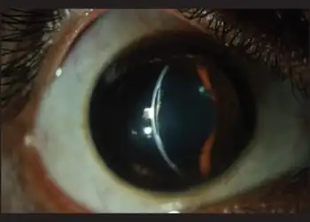 | |
| Keratoglobus with healed hydrops ( tear in the Descemet's membrane is visible) | |
 | |
| Specialty | Ophthalmology |
Keratoglobus (from Greek: kerato- horn, cornea; and Latin: globus round) is a degenerative non-inflammatory disorder of the eye in which structural changes within the cornea cause it to become extremely thin and change to a more globular shape than its normal gradual curve. It causes corneal thinning, primarily at the margins, resulting in a spherical, slightly enlarged eye.
It is sometimes equated with "megalocornea".[1]
Signs and symptoms
The presentation of Keratoglobus is consistent with the following:[2]
- Myopia
- Globular protrusion of cornea
Pathophysiology
Keratoglobus is a little-understood disease with an uncertain cause, and its progression following diagnosis is unpredictable. If afflicting both eyes, the deterioration in vision can affect the patient's ability to drive a car or read normal print. It does not however lead to blindness.
Diagnosis
In terms of the diagnosis of keratoglobus is done via the clinical presentation and characteristic findings.[3]
Treatment
Treatment includes the use of protective eye glasses.[3] A number of surgical options are also available.[3]
Further progression of the disease usually leads to a need for corneal transplantation because of extreme thinning of the cornea. Primarily, large size penetrating keratoplasty has been advocated. Recent additions of techniques specifically for keratoglobus include the "tuck procedure",[4] whereby a 12 mm corneo-scleral donor graft is taken and trimmed at its outer edges. A host pocket is formed at the limbal margin and the donor tissue is "tucked" into the host pocket.
Prognosis
Keratoglobus continues to be a somewhat mysterious disease, but it can be successfully managed with a variety of clinical and surgical techniques. The patient is at risk for globe perforation because the thinned out cornea is extremely weak.
Epidemiology
It is a much rarer condition than keratoconus, which is the most common dystrophy of the cornea.[5] Similar to keratoconus it is typically diagnosed in the patient's adolescent years and attains its most severe state in the twenties and thirties.
References
- ↑ "keratoglobus" at Dorland's Medical Dictionary
- ↑ "Keratoglobus - EyeWiki". eyewiki.aao.org. Archived from the original on 8 June 2023. Retrieved 17 August 2023.
- 1 2 3 Wallang, BS; Das, S (September 2013). "Keratoglobus". Eye. London. 27 (9): 1004–1212. doi:10.1038/eye.2013.130. PMC 3772364. PMID 23807384.
- ↑ Kaushal S, Jhanji V, Sharma N, Tandon R, Titiyal JS, Vajpayee RB (February 2008). ""Tuck In" Lamellar Keratoplasty (TILK) for corneal ectasias involving corneal periphery". Br J Ophthalmol. 92 (2): 286–290. doi:10.1136/bjo.2007.124628. PMID 18227208. S2CID 36981311.
- ↑ "Review of Optometry". Archived from the original on 2007-03-10. Retrieved 2006-10-17.
External links
| Classification |
|---|